Tissue level of organisation
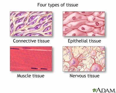
In the article “tissue level of organisation”, we will explore the fundamental building block of the human body – the tissue. Tissues are collections of similar cells that work together to perform specific functions. We will discuss the four main types of human tissues – epithelial, connective, muscle, and nervous – and delve into their unique structures, location and functions.
Epithelial tissue
Epithelial tissue, also known as epithelium, is one of the four main types of tissues in the human body. It forms a continuous layer of cells that covers the outer surface of the skin and lines the body cavities. This tissue plays a crucial role in various functions such as absorption, protection, sensation, and secretion.
Structure of Epithelial Tissue
- Epithelial tissue is composed of a tightly fitted continuous layer of cells.
- One surface of the tissue is exposed to either the external environment or the body fluid.
- The other surface is attached to the tissue by a membrane, which consists of fibers and polysaccharides secreted by the epithelial cells.
- There are specialized junctions present between the cells of the epithelium that link individual cells.
- These junctions include tight junctions that prevent leakage across tissues, adhering junctions that keep the neighboring tissues well cemented together, and gap junctions that facilitate the movement of ions and molecules across the tissue.
Epithelial cells
- The building blocks of epithelial tissue are the epithelial cells.
- These cells are tightly packed together, forming a ‘cell-rich’ tissue with very little extracellular space between them.
- They form uniform sheets of cells, often in layers.
- Epithelial cells are characterized by the presence of a large nucleus and a polar arrangement.
- That is, they have a defined structural orientation, with the membranes on either side possessing a distinct set of proteins.
- Some types of cells, including some epithelial cells, have characteristics on the surface of the cell that help them perform certain functions, including microvilli and cilia.
Types of Epithelial Tissue
There are several different types of epithelial cells based on their shape and arrangement.
Simple Epithelia
This is a single layer of cells. It includes:
- Simple Squamous: Thin and flat cells, like fish scales.
- Simple Cuboidal: Cells that are roughly square or round in shape.
- Simple Columnar: Elongated cells involved in absorption of materials.
Stratified Epithelia
This consists of two or more layers of cells. It includes:
- Stratified Squamous: Multiple layers of flat cells.
- Stratified Cuboidal: Multiple layers of square or round cells.
- Stratified Columnar: Multiple layers of elongated cells.
Pseudostratified Columnar
This appears to have multiple layers due to the irregular placement of the nuclei within the cells, but each cell is in contact with the basement membrane.
Transitional (or Urothelium)
This type of epithelium can stretch and expand. It is found in the urinary bladder and parts of the urinary tract
Location of Epithelial Tissue
- Epithelial tissue forms the outer covering of the skin and also lines the body cavity.
- It forms the lining of various tracts in the body, including the respiratory, digestive, reproductive, and excretory tracts.
- It also lines the outer surfaces of organs and blood vessels throughout the body, as well as the inner surfaces of cavities in many internal organs.
Functions of Epithelial Tissue
- Secretion: Epithelial cells are often specialized to secrete specific substances such as hormones, enzymes, or other fluids.
- Protection: As it covers the entire body surface, it serves as the first line of defense against any kind of mechanical injury, chemical exposure, excessive fluid loss, and infections.
- Absorption: Certain types of epithelial cells, such as those found in the intestines, are specialized for absorbing nutrients from the food we eat.
- Transportation: It plays a crucial role in the transportation of substances that the epithelial cell needs to internalize or expel.
- Sensation: Some epithelial cells have sensors that are receptors. They are capable of picking up signals and transporting them. For instance, when you touch a soft piece of bread, the sensors detect corresponding signals from your hands. Then, they can send the signal to the brain.
Connective tissue
Connective tissue is one of the four basic types of animal tissue. It is found throughout the body, serving to connect and support other tissues and organs. It is composed of various types of cells and extracellular fibers in a ground substance.
Structure of connective tissue
Connective tissues are composed of three main components: cells, fibers, and extracellular matrix.
Cells
The cells in connective tissue can be divided into fixed cells (which remain within the tissue) and wandering cells (which move through and can leave the tissue). Fixed cells include fibroblasts, adipocytes, and mesenchymal cells, while wandering cells include leukocytes.
Fibers
There are three types of fibers found within connective tissue: collagen fibers, elastic fibers, and reticular fibers.
- Collagen Fibers: These are the most widespread fibers in connective tissue and are made up of the fibrous protein, collagen. Collagen fibers are flexible and have high tensile strength, comparable to steel.
- Elastic Fibers: These fibers form a network and can be stretched like a rubber band. They are made up of the protein elastin and are able to retain their original shape and size once the force is removed.
- Reticular Fibers: These consist of collagen and glycoproteins. They are thin and form a delicate network. Reticular fibers join connective tissues to neighboring tissues.
Extracellular Matrix
This is a gel-like substance surrounding the cells and fibers. It is composed of water, proteins, and polysaccharides
Types of Connective Tissue
- Loose Connective Tissue: This type of tissue includes areolar tissue, reticular tissue, and adipose tissue. It acts as a padding under the skin and elsewhere.
- Dense Connective Tissue: This tissue is composed of large amounts of closely packed collagenous fibers. It includes dense regular, dense irregular, and elastic connective tissues.
- Specialized Connective Tissue: This category includes cartilage, bone, and blood. Each has a unique structure and function.
Location of Connective Tissue
- Connective tissues are widely distributed in every part of the body.
- They are found in between other tissues everywhere in the body, including the nervous system.
- They are most abundant in the reticular tissue of soft organs, such as liver and spleen, where they anchor and provide structural support to the parenchyma.
- They are also found in the external ear, larynx, joint surfaces and growth zone of bones, rings around the trachea, between ribs and sternum, intervertebral discs, etc.
Functions of Connective Tissue
- Binding and Support: Connective tissue can bind together and support various tissues and organs in the body.
- Protection: Certain types of connective tissue, such as bone and cartilage, protect vital organs from injury.
- Insulation: Adipose tissue, a type of connective tissue, insulates the body and helps maintain body temperature.
- Transportation: Blood, another type of connective tissue, is responsible for transporting oxygen, nutrients, and other substances throughout the body.
Muscular tissue
Muscular tissue is a specialized tissue found in animals which functions by contracting, thereby applying forces to different parts of the body. It is made up of thin and elongated cells called muscle fibers. The cytoplasm in the muscle fibers is called sarcoplasm. It contains a network of membrane called the sarcoplasmic reticulum.
Structure of Muscular Tissue
- Muscular tissues are composed of thin and elongated cells called muscle fibers.
- The cytoplasm in the muscle fibers is called sarcoplasm, which contains a network of membranes known as the sarcoplasmic reticulum.
- The membrane surrounding the muscle fibers is called sarcolemma.
- Muscular tissues are bundled together and surrounded by a tough connective tissue similar to cartilage known as epimysium.
- The bundle of nerve cells that run in long fibers are called fascicles, which are surrounded by the epimysium.
- The fascicles are surrounded by a protective layer known as perimysium.
- Another protective layer, the endomysium, surrounds the fibers.
- These layers and muscles help in the contraction of different parts of the muscles.
Types of Muscular Tissue
There are three types of muscular tissue: Skeletal Muscle Tissue, Smooth Muscle Tissue, and Cardiac Muscle Tissue.
Skeletal Muscle Tissue
These muscles are attached to the skeleton and help in its movement. These muscles are also known as striated muscles because of the presence of alternate patterns of light and dark bands. These light and dark bands are sarcomeres which are highly organized structures of actin, myosin, and proteins. These add to the contractility and extensibility of the muscles. Skeletal muscles are voluntary muscles composed of muscle fibers.
Smooth Muscle Tissue
These are non-striated, involuntary muscles controlled by the Autonomous Nervous System. It stimulates the contractility of the digestive, urinary, reproductive systems, blood vessels, and airways. The actin and myosin filaments are very thin and arranged randomly, hence no striations.
Cardiac Muscle Tissue
This type of muscle tissue is found in the heart. It is striated like skeletal muscle but, unlike skeletal muscle, it can’t be controlled voluntarily.
Location of Muscular Tissue
- Muscular tissues are widely distributed in every part of the body.
- They are found in between other tissues everywhere in the body, including the nervous system.
- They are most abundant in the reticular tissue of soft organs, such as liver and spleen, where they anchor and provide structural support to the parenchyma.
- They are also found in the external ear, larynx, joint surfaces and growth zone of bones, rings around the trachea, between ribs and sternum, intervertebral discs, etc.
Functions of Muscular Tissue
- Muscular tissue functions as a single unit, and is often connected to the same nerve bundles.
- A nerve impulse traveling from the brain or another outside signal tells the muscle to contract.
- The nerve impulse is transferred almost instantaneously to all the nerve cells in the muscle tissue, and the entire muscle contracts.
- At the cellular level, each muscle cell has a complex of proteins containing actin and myosin.
- These proteins slide past one another when the signal to contract is received.
- The filaments are connected to the ends of the cells, and as they slide past one another, the cell contracts in length.
- A single cell can contract up to 70% in length, which shortens the entire muscle when contraction happens.
- Muscle tissue can be used to move bones, compress chambers, or squeeze various organs.
Nervous tissue
Nervous tissue, also known as neural tissue, is the main tissue component of the nervous system. It monitors and regulates the functions of the body. Nervous tissue consists of two types of cells: nerve cells or neurons and glial cells. Neurons transmit nerve impulses and glial cells help transmit these impulses and also provide nutrients to neurons.
Structure of nervous tissue
- Nervous tissue is made up of nerve cells or neurons, all of which consist of an axon.
- Axons are long stem-like projections emerging out of the cell, responsible for communicating with other cells called the target cells, thereby passing impulses.
- The main part of the neuron is the cell body which contains the nucleus, cytoplasm, and cell organelles.
- Extensions of the cell membrane are referred to as processes.
- Dendrites are highly branched processes, responsible for receiving information from other neurons and synapses (specialized point of contact).
- Information in a neuron is unidirectional as it passes through neurons from dendrites, across the cell body down the axon.
Types of nervous tissue
- Nervous tissue is grouped into two main categories: neurons and neuroglia.
- Neurons, or nerves, transmit electrical impulses.
- Neuroglia do not transmit impulses; instead, they have many other functions including supporting and protecting neurons.
- There are four types of neuroglia found in the CNS: astrocytes, microglial cells, ependymal cells, and oligodendrocytes.
- Two types of neuroglia found in the PNS are satellite glial cells and Schwann cells.
Neurons
- Neurons, also known as nerve cells, are the fundamental units of the nervous system.
- They are specialized cells that transmit signals called nerve impulses, or action potentials.
- An action potential is a quick rise and fall in the electrical membrane potential of the neuron, which transmits signals from one neuron to the next.
Neuroglia
Neuroglia, also known as glial cells, are non-neuronal cells that maintain homeostasis, form myelin, and provide support and protection for neurons in the central nervous system (CNS) and peripheral nervous system (PNS). There are four types of neuroglia found in the CNS: astrocytes, microglial cells, ependymal cells, and oligodendrocytes.
- Astrocytes: These are star-shaped cells that provide physical and nutritional support for neurons.
- Microglial Cells: These are small cells that remove waste and pathogens by phagocytosis.
- Ependymal Cells: These cells line the cavities of the CNS and secrete cerebrospinal fluid.
- Oligodendrocytes: These cells provide support and insulation to axons in the CNS by forming a myelin sheath.
Two types of neuroglia found in the PNS are satellite glial cells and Schwann cells.
- Satellite Glial Cells: These cells surround neuron cell bodies within ganglia.
- Schwann Cells: Also known as neurolemmocytes, these cells form the myelin sheath around axons in the PNS.
Location of Nervous Tissue
- The nervous tissue is located throughout the body in the peripheral nerves as well as in the organs of the central nervous system such as the spinal cord and brain.
- The nerve tissue or the nervous tissue is the chief tissue component of the two major parts of the nervous tissue – Central nervous system (CNS) formed by the spinal cord and the brain, and the branching peripheral nerves of the peripheral nervous system (PNS) that control and regulate the functions of the body and their activities.
Functions of Nervous Tissue
- Nervous tissue makes up for the CNS and PNS of the nervous system.
- It contains two distinct cells – neurons and glial cells.
- Neurons secrete chemical neurotransmitters which are responsible for stimulating other neurons as a result of a stimuli.
- Nervous tissue has a role in controlling bodily functions such as digestion.
- It sends and carries signals to and from the different parts of the body.
- It controls the body’s movements.

Conclusion
In conclusion, human tissues are fundamental to the structure and function of our bodies. They are categorized into four main types: epithelial, connective, muscular, and nervous tissues. Each type has a unique structure and function that contributes to our overall health and well-being.
Epithelial tissue forms the outer layer of our skin and internal organs, providing protection and allowing for the absorption and secretion of substances. Connective tissue, on the other hand, provides support and connects different tissues and organs of the body. It is composed of various types of cells and extracellular fibers in a ground substance.
Muscular tissue, which is responsible for movement and force exertion, is made up of specialized cells called muscle fibers. These fibers contract and relax to facilitate movement. Lastly, nervous tissue, the main component of our nervous system, plays a crucial role in transmitting signals across the body, thereby regulating bodily functions.
Understanding these tissues and their functions is fundamental to the study of human biology and medicine.
For more regular updates you can visit our social media accounts,
Instagram: Follow us
Facebook: Follow us
WhatsApp: Join us
1 thought on “Tissue Level of Organisation”