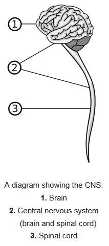
Central nervous system
The central nervous system is like the boss of our body. It has two main parts: the brain and the spinal cord. The brain is where we think and feel, and the spinal cord is like a big cable that sends messages between the brain and the body. It’s really important because it helps us do everything from moving our hands to remembering our favorite song. We’re going to learn all about how it works and why it’s so special.
Meninges
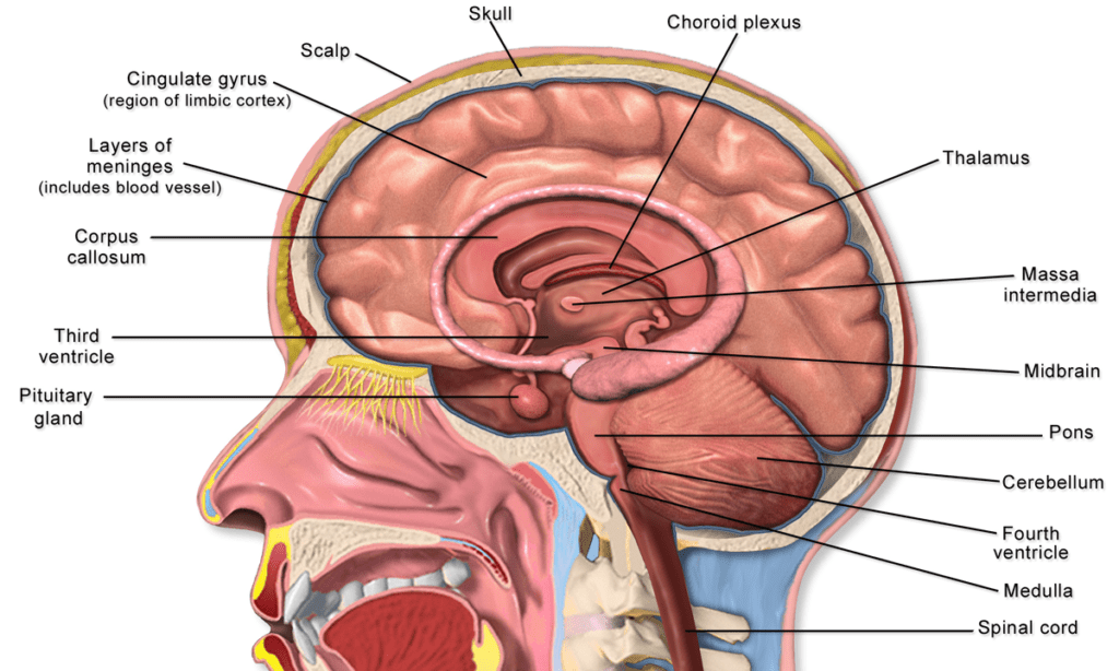
The meninges are three protective layers of membranes that cover and safeguard your brain and spinal cord (collectively known as the central nervous system or CNS).
Anatomy of the Meninges
Dura Mater
- The outermost layer, akin to a sturdy shield.
- In Latin, “dura mater” translates to “hard mother.”
- Comprises two layers of tough connective tissue:
- Periosteal cranial dura: The outer layer attached to the skull.
- Meningeal dura: The middle layer.
- In the spine, it consists solely of the meningeal layer.
- Contains dural venous sinuses, allowing blood to exit the brain and permitting cerebrospinal fluid (CSF) reabsorption.
Arachnoid Mater
- The middle layer, like a delicate spider web.
- Lies between the dura mater and the innermost layer (pia mater).
- Forms a protective barrier around the central nervous system (CNS).
- The subarachnoid space beneath it houses CSF.
Pia Mater
- The innermost layer, intimately embracing brain tissue.
- Together with the arachnoid mater, it constitutes the leptomeninges.
Functions of the Meninges
- Protection: Acts as a shock absorber, shielding the CNS from trauma (such as head injuries). Prevents excessive movement of the brain.
- Anchoring: Keeps the brain in place, preventing it from jostling too much.
- Support System: Supports blood vessels (including the middle meningeal artery). Facilitates CSF flow, which cushions and protects the brain and spinal cord.
Ventricles of brain
Anatomy of Brain Ventricles
First and second ventricles (Lateral Ventricles)
Location: Situated within each cerebral hemisphere.
Structure
- Body: The central part.
- Anterior (Frontal) Horn: Extends into the frontal lobe.
- Posterior (Occipital) Horn: Reaches into the occipital lobe.
- Inferior (Temporal) Horn: Extends into the temporal lobe.
Choroid Plexus: Each lateral ventricle houses a choroid plexus, responsible for synthesizing cerebrospinal fluid (CSF).
Third Ventricle
Location: Found in the diencephalon (between the thalamus and hypothalamus).
Notable Features
- Supraoptic Recess: Superior to the optic chiasma.
- Infundibular Recess: Superior to the pituitary infundibulum.
- Suprapineal Recess: Superior to the pineal gland.
- Pineal Recess: Protrudes into the pineal infundibulum.
Choroid Plexus of Third Ventricle: Responsible for CSF production.
Fourth Ventricle
Location: Nestled within the brainstem.
Floor: Forms a rhomboid fossa.
Roof: Comprises the superior and inferior medullary vela of the cerebellum.
Cisterns Associated with Fourth Ventricle
- Suprasellar (Chiasmatic) Cistern
- Interpeduncular Cistern
- Prepontine Cistern
- Cistern of the Corpus Callosum
Foramina Connecting Ventricles
- Interventricular Foramina (Monro): Link lateral ventricles to the third ventricle.
- Cerebral Aqueduct (Sylvius): Connects the third ventricle to the fourth ventricle.
- Median Aperture (Magendie): Connects the fourth ventricle to the subarachnoid space.
- Right & Left Lateral Apertures (Luschka): Also connect the fourth ventricle to the subarachnoid space.
Functions of Brain Ventricles
CSF Production and Circulation
- Choroid plexuses within ventricles synthesize CSF.
- CSF circulates through the ventricles and subarachnoid space, bathing the brain and spinal cord.
Protection and Cushioning
- CSF acts as a cushion, protecting the brain from mechanical shocks.
- It also helps maintain a stable environment for neural function.
Cerebrospinal fluid
Anatomy of Cerebrospinal Fluid (CSF)
What Is CSF?
- CSF is a clear, colorless plasma-like fluid that bathes the central nervous system (CNS).
- It circulates through a system of cavities within the brain and spinal cord:
- Ventricles: Located within the brain.
- Subarachnoid space: Surrounding the brain and spinal cord.
- Central canal of the spinal cord.
Choroid Plexus
Specialized tissue responsible for producing CSF. Found within the;
- Lateral ventricles
- Third ventricle
- Fourth ventricle
CSF secretion equals its removal, maintaining a constant volume of around 150-270 milliliters within the CNS.
Functions of CSF
- Cushioning and Protection: Acts as a shock absorber, safeguarding the brain and spinal cord from mechanical force (e.g., impacts).
- Immunological Protection: Provides basic immune defense to the CNS.
- Waste Removal: Clears metabolic waste products.
- Transport of Neuromodulators and Neurotransmitters: Facilitates communication within the nervous system.
Brain
Structure
Cerebrum:
- The largest part of the brain.
- Composed of two cerebral hemispheres (left and right). Each hemisphere has:
- An inner core of white matter.
- An outer surface called the cerebral cortex, made of grey matter.
The cortex includes
- Neocortex: Six neuronal layers.
- Allocortex: Three or four layers.
Divided into four lobes
- Frontal Lobe: Associated with executive functions (self-control, planning, reasoning) and abstract thought.
- Temporal Lobe: Involved in memory, hearing, and language comprehension.
- Parietal Lobe: Processes sensory information (touch, temperature, pain).
- Occipital Lobe: Dedicated to vision.
Functions of the Cerebrum
Sensory Processing:
- Receives and interprets sensory input from our environment.
- Different regions specialize in vision, hearing, touch, taste, and smell.
Motor Control
- Initiates voluntary movements.
- Motor areas map to specific body parts (motor homunculus).
Language and Communication
- Broca’s area (left hemisphere) governs speech production.
- Wernicke’s area (left hemisphere) is involved in language comprehension.
Memory and Learning
- Hippocampus (within the temporal lobe) plays a key role.
Emotions and Behavior
- Frontal lobe influences personality, decision-making, and social behavior.
Higher Cognitive Functions
- Reasoning, problem-solving, creativity, and abstract thinking.
Brainstem
Connects the cerebrum to the spinal cord. It comprises,
- Midbrain: Regulates visual and auditory reflexes.
- Pons: Controls breathing, swallowing, and facial movements.
- Medulla Oblongata: Vital for heart rate, blood pressure, and respiration.
Functions of the Brainstem
Vital Body Functions
- The brainstem sends messages back and forth between the brain and other body parts.
- It regulates many involuntary actions, such as:
- Balance
- Blood pressure
- Breathing
- Eye movements
- Facial movements and sensations
- Hearing
- Heart rate
- Sleep and wakefulness
- Swallowing
- Taste
Brainstem Reflexes
These are immediate and involuntary motor responses that help us survive and adapt to changes in our environment.
- Cardiovascular reflexes: Regulating heartbeat and blood pressure.
- Gag reflex: Protecting our airways.
- Swallowing reflex: Moving food and liquids from the mouth to the stomach.
- Pupillary light reflex: Adjusting pupil size based on lighting changes.
- Vestibulo-ocular reflex: Stabilizing our eyes during head or body movements.
- Respiratory reflexes: Regulating breathing, coughing, and sneezing.
Cerebellum
- Located at the back of the brain.
- Responsible for balance, coordination, and fine motor control.
- Connected to the brainstem via cerebellar peduncles.
- Its name comes from Latin, meaning “little brain.”
- The cerebellum has an ovoid shape, slightly flattened, and a central recess.
Functions of the Cerebellum
Motor Coordination
- The cerebellum fine-tunes muscle movements, ensuring smooth coordination.
- It adjusts muscle tone, posture, and balance.
Precision and Timing
- When you reach for an object, type on a keyboard, or play a musical instrument, the cerebellum ensures precise timing and accuracy.
Learning and Adaptation
- It plays a role in motor learning, allowing you to acquire new skills and refine existing ones.
Emotional Regulation
- Recent research suggests that the cerebellum is involved in emotional processing and decision-making.
Ventricular System
- Four interconnected ventricles within the cerebrum.
- Cerebrospinal fluid (CSF) is produced and circulated here.
- Ventricles include the lateral ventricles, third ventricle, and fourth ventricle.
Subcortical Structures (Beneath the Cortex)
- Thalamus: Relays sensory and motor signals.
- Hypothalamus: Regulates body temperature, hunger, thirst, and hormones.
- Limbic System: Involved in emotions, memory, and motivation (includes the amygdalae and hippocampi).
- Basal Ganglia: Influences movement and cognition.
- Brainstem Structures: Epithalamus, pineal gland, pituitary gland, and more.
Functions of brain
Sensory Processing
- The brain receives information from your five senses: sight, smell, sound, touch, and taste.
- It also processes inputs like touch, vibration, pain, and temperature from the rest of your body.
- By interpreting this information, your brain helps you understand and associate meaning with your surroundings.
Thoughts and Decisions
- The brain enables complex cognitive processes, including reasoning, problem-solving, and decision-making.
- It allows you to weigh options, plan, and choose appropriate actions.
Memories and Emotions
- The brain stores memories, both short-term and long-term.
- It processes emotions, influencing how you feel and react to various situations.
Motor Function and Coordination
- The brain controls voluntary movements, allowing you to walk, talk, write, and perform intricate tasks.
- It maintains balance, coordination, and fine motor skills through structures like the cerebellum.
Perception of Sensations
- The brain processes sensory input, including pain, temperature, pressure, and proprioception (awareness of body position).
- It constructs your perception of the world around you.
Automatic Behaviors: The brain regulates essential functions without conscious effort:
- Breathing
- Heart rate
- Sleep and wake cycles
- Digestion
- Temperature control
Speech and Language Functions
- Specific brain areas handle language comprehension, speech production, and communication.
Fight or Flight Response (Stress Response)
- When faced with danger or stress, the brain triggers physiological changes to prepare your body for action.
Spinal cord
Structure

Location and Protection
- The spinal cord runs through the vertebral column (also known as the spine).
- It begins at the end of the brainstem and extends down almost to the bottom of the spine.
- The vertebral column’s vertebrae (bones) surround and protect the spinal cord.
Tissues and Layers
- Like your brain, the spinal cord is covered by protective layers called meninges:
- Dura Mater: The tough outer layer that shields the spinal cord from injury.
- Arachnoid Mater: The middle layer between the epidural and subarachnoid space.
- Pia Mater: The innermost layer that directly covers the spinal cord.
Segments and Regions: The spinal cord is divided into three main regions;
- Cervical (neck): Located in the neck area.
- Thoracic (chest): Found in the chest region.
- Lumbar (lower back): Situated in the lower back.
Afferent Nerve Tracts (Sensory Pathways)
Afferent neurons, also known as sensory neurons, carry information from sensory receptors (such as those in the skin, eyes, ears, nose, and tongue) to the central nervous system (CNS)—specifically, to the brain and spinal cord. These neurons allow us to sense, perceive, and react to stimuli from the outside world.
Functions
Sensory Information: Afferent neurons transmit sensory information related to;
- Vision
- Hearing
- Smell
- Taste
- Touch
- Pain
- Temperature
When you touch a hot pan, for example, afferent neurons send information about that stimulus up the spinal cord to the brain.
Processing and Response
- The brain processes this sensory input and decides how to respond.
- Association neurons (interneurons) relay information between afferent and efferent neurons.
- Ultimately, motor efferent neurons communicate with muscles, indicating what motion to perform (e.g., pulling away from the hot pan).
Efferent Nerve Tracts (Motor Pathways)
- Efferent neurons, also called motor neurons, carry signals from the brain to the peripheral nervous system.
- They initiate actions by relaying commands to muscles and glands.
Functions
Motor Control: Efferent neurons control voluntary movements;
- Walking
- Reaching
- Writing
- Speaking
- They also regulate involuntary processes like heart rate and digestion.
Response to Sensory Input
- When you decide to pull your hand away from the hot pan, efferent neurons transmit the motor command to the muscles.
Reflex activity
What Is a Reflex?
- A reflex action is an automatic, nearly instantaneous movement or response to a stimulus.
- Unlike voluntary actions, reflexes do not involve conscious decision-making or the brain’s direct involvement.
- Instead, the spinal cord coordinates the response, allowing the impulse to reach the effector (muscles or glands) quickly.
Components of a Reflex Arc
Stimulus
- The event or condition that triggers the reflex.
- Examples: Touching a hot surface, sudden loud noise, or a puff of air in your eye.
Receptor
- Specialized sensory structures (such as nerve endings) that detect the stimulus.
- Located in the skin, muscles, tendons, or organs.
Sensory Neuron (Afferent Pathway)
- Carries the sensory information from the receptor to the spinal cord or brainstem.
- The impulse travels along the afferent nerve fibers.
Integration Center (Spinal Cord or Brainstem)
- The reflex arc’s central processing unit.
- Determines the appropriate response based on the sensory input.
- Involves interneurons (relay neurons) within the spinal cord.
Motor Neuron (Efferent Pathway)
- Carries the response signal from the integration center to the effector (muscles or glands).
- The impulse travels along the efferent nerve fibers.
Effector
- The muscle or gland that carries out the response.
- Examples: Contracting a muscle (as in the knee-jerk reflex) or secreting saliva (as in the salivary reflex).
Examples of Reflex Actions
Knee-Jerk Reflex
- When a doctor taps your knee with a rubber hammer, your leg automatically kicks forward.
- Involves the quadriceps muscle and the patellar tendon.
Withdrawal Reflex
- Touching a hot surface causes you to pull your hand away instantly.
- Protects you from potential harm.
Pupillary Light Reflex
- When exposed to bright light, your pupils constrict to reduce the amount of light entering your eyes.
Conclusion
The Central Nervous System (CNS) stands as the epicenter of our neural universe. Comprising the brain and spinal cord, it orchestrates our every thought, emotion, and bodily function. The brain, with its cerebral hemispheres, cerebellum, and brainstem, processes sensory input, controls motor functions, and interprets sensations. The spinal cord, encased in protective layers, connects the brain to the rest of the body, relaying messages and coordinating reflexes. Whether it’s the rapid knee-jerk reflex or the intricate dance of voluntary movements, the CNS conducts this neural symphony—a marvel to behold!
For more regular updates you can visit our social media accounts,
Instagram: Follow us
Facebook: Follow us
WhatsApp: Join us
Telegram: Join us
1 thought on “Central Nervous System”