Joints
“Joints, also known as articulations, are the fascinating intersections in the human body where bones meet. They are the unsung heroes of our daily lives, enabling us to perform a myriad of movements, from simple gestures like waving hello to complex physical activities like gymnastics or martial arts. The human body, with its 206 bones, is a marvel of joint engineering. Each bone (except the hyoid bone in the neck) is connected to at least one other bone, forming a complex network of joints that work together in harmony. Joints are not all created equal. They are classified both structurally and functionally, providing a comprehensive understanding of their diverse nature.
Classification of joints
Structurally, joints are categorized based on the type of connective tissue that binds the bones together. Some joints are bound by fibrous connective tissue, others by cartilage, and some articulate within a fluid-filled space called a joint cavity.
Functionally, joints are classified based on the degree of movement they allow. Some joints are immobile, some have limited mobility, and others are freely movable. This functional diversity allows us to perform a wide range of actions, from the rigidity required for standing to the flexibility needed for running or dancing.
Structural classification of joints
Structurally, joints are classified based on the type of connective tissue that binds the bones together. There are three main types of structural classifications:
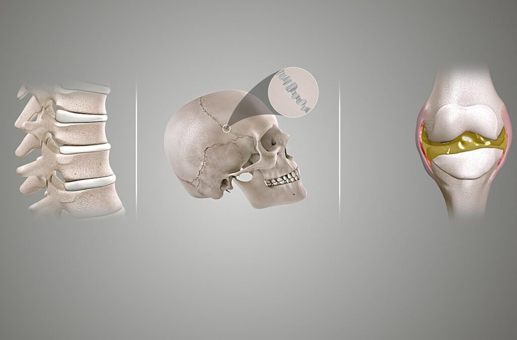
Fibrous Joints
These are the joints where the adjacent bones are united by fibrous connective tissue. They have no joint cavity and are connected via fibrous connective tissue. The skull bones are held together by fibrous joints.
Characteristics of Fibrous Joints
- Direct Connection: At a fibrous joint, the adjacent bones are directly connected to each other by fibrous connective tissue.
- No Joint Cavity: Fibrous joints do not have a joint cavity between them.
- Fibrous Connective Tissue: The bones at fibrous joints are connected by dense connective tissue consisting mainly of collagen.
- Immovable: Fibrous joints are also called “fixed” or “immovable” joints because they do not move.
- Types of Fibrous Joints: There are three types of fibrous joints – sutures, syndesmoses, and gomphoses.
Functions of Fibrous Joints
- Protection: Fibrous joints strongly unite adjacent bones and thus serve to provide protection for internal organs.
- Strength and Stability: These joints typically require strength and stability over a range of movement.
- Sutures: Sutures are the fibrous joints that hold the plates of your skull together. They help protect the brain and form the face.
- Syndesmoses: Syndesmoses are joints that hold two closely related bones together in place. An example is the joint between the shaft regions of the long bones in the forearm and in the leg.
- Gomphoses: Gomphoses are the joints that anchor a tooth to its socket in the jaw. They hold your teeth in place in your jaw bones.
Cartilaginous Joints
At these joints, the bones are joined by hyaline cartilage or fibrocartilage. They also have no joint cavity. An example of a cartilaginous joint is the joint between the manubrium and the sternum.
Characteristics of Cartilaginous Joints
- Cartilage Connection: Cartilaginous joints are connected entirely by cartilage, either fibrocartilage or hyaline.
- No Joint Cavity: These types of joints lack a joint cavity.
- Limited Movement: Cartilaginous joints allow more movement between bones than a fibrous joint but less than the highly mobile synovial joint.
- Types of Cartilaginous Joints: There are two types of cartilaginous joints – synchondroses and symphyses.
Functions of Cartilaginous Joints
- Shock Absorption: Cartilage is a strong, flexible connective tissue that acts as a shock absorber throughout your body.
- Reduced Friction: Cartilage at the end of your bones reduces friction and prevents them from rubbing together when you use your joints.
- Synchondroses: A synchondrosis is a cartilaginous joint where bones are joined together by hyaline cartilage, or where bone is united to hyaline cartilage. An example is the epiphyseal plate (growth plate) of a growing long bone.
- Symphyses: A symphysis is a type of cartilaginous joint where the bones are joined by fibrocartilage. An example is the pubic symphysis, where the pubic portions of the right and left hip bones of the pelvis are joined together by fibrocartilage.
Synovial Joints
These are the most common type of joints in the body. At a synovial joint, the articulating surfaces of the bones are not directly connected, but instead come into contact with each other within a joint cavity that is filled with a lubricating fluid. This fluid-filled space allows for free movement between the bones. Examples of synovial joints include the shoulder, elbow, knee, and hip joints.
Characteristics of Synovial Joints
- Joint Cavity: A key structural characteristic for a synovial joint is the presence of a joint cavity. This fluid-filled space is the site at which the articulating surfaces of the bones contact each other.
- Articular Cartilage: The articulating surfaces of the bones are covered by a thin layer of hyaline cartilage. This acts like a coating over the bone surface, allowing the articulating bones to move smoothly against each other without damaging the underlying bone tissue.
- Synovial Membrane: Lining the inner surface of the articular capsule is a thin synovial membrane. The cells of this membrane secrete synovial fluid.
- Ligaments: Outside of their articulating surfaces, the bones are connected together by ligaments, which are strong bands of fibrous connective tissue.
- Articular Capsule: The walls of the joint cavity are formed by the articular capsule, a fibrous connective tissue structure that is attached to each bone just outside the area of the bone’s articulating surface.
Functions of Synovial Joints
- Movement: Synovial joints allow for smooth movements between the adjacent bones. The ability of the bones to move smoothly against each other within the joint cavity, and the freedom of joint movement this provides, means that each synovial joint is functionally classified as a diarthrosis.
- Lubrication: A very thin layer of slippery, viscous joint fluid, called synovial fluid, separates and lubricates the two cartilage-covered bone surfaces.
- Nourishment: The synovial fluid also provides nourishment to the articular cartilage, which does not contain blood vessels.
- Support: Ligaments strengthen and support the joint by anchoring the bones together and preventing their separation.
Functional classification of joints
Synarthrosis: These are immovable joints. They are rigid and provide stability to the skeletal structure. Examples include:
- Sutures: Found in the skull, they are fibrous joints that fuse bones together.
- Gomphosis: A type of joint that anchors a tooth to its socket in the jaw.
Amphiarthrosis: These joints allow for a small amount of movement and are more flexible than synarthrosis but less so than diarthrosis. Examples include:
- Syndesmosis: A joint where bones are connected by a ligament, such as the distal joint between the tibia and fibula.
- Symphysis: A joint where bones are connected by fibrocartilage, like the pubic symphysis in the pelvis.
Diarthrosis (Synovial Joints): These are freely movable joints that allow for a wide range of motion. They are characterized by a joint cavity filled with synovial fluid, which lubricates the joint. Examples include:
- Ball-and-socket joints: Such as the shoulder and hip joints, which allow for rotational movement and a wide range of motion.
- Hinge joints: Like the elbow and knee, which allow for bending and straightening motions.
- Pivot joints: Such as the atlantoaxial joint in the neck, which allows for rotational movement.
- Gliding joints: Found between the small bones of the wrist and foot, allowing sliding movements.
- Saddle joints: Such as the thumb joint, which allows for grasping and rotation.
- Condyloid joints: Like the wrist joint, allowing movement but no rotation
Types of joint movement
Gliding Movements: These occur at flat bone surfaces where the bones slide over each other. An example is the movement at the carpals of the wrist.
Flexion and Extension: These movements are in the sagittal (anterior–posterior) plane of motion. Flexion decreases the angle between two body parts, while extension increases it. These movements take place at the shoulder, hip, elbow, knee, wrist, metacarpophalangeal, metatarsophalangeal, and interphalangeal joints.
Abduction and Adduction: These movements are in the coronal (medial–lateral) plane of movement. Abduction is the movement away from the midline of the body, while adduction is the movement towards the midline. These movements occur at the shoulder, hip, wrist, metacarpophalangeal, and metatarsophalangeal joints.
Circumduction: This is a circular movement of a body part, using the sequential combination of flexion, adduction, extension, and abduction motions. This movement occurs at ball-and-socket joints like the shoulder and hip.
Rotation: This involves turning of the head side to side or twisting of the body. Medial and lateral rotation of the upper limb at the shoulder or lower limb at the hip involves turning the anterior surface of the limb toward the midline of the body (medial or internal rotation) or away from the midline (lateral or external rotation).
Pronation and Supination: These are unique to the forearm. Pronation is the inward roll of the forearm, while supination is the outward roll of the forearm.
Dorsiflexion and Plantar Flexion: These movements are unique to the foot. Dorsiflexion is the bending of the foot upwards, while plantar flexion is the bending of the foot downwards.
Conclusion
Joints, or articulations, are the fundamental components of the human body that facilitate movement and provide stability. They are classified structurally into fibrous, cartilaginous, and synovial joints, each with unique characteristics and functions. Fibrous joints, connected by dense fibrous tissue, provide strength and stability. Cartilaginous joints, connected by cartilage, offer a balance between stability and mobility. Synovial joints, characterized by a fluid-filled joint cavity, allow for a wide range of movements.
The movements at these joints, including flexion, extension, abduction, adduction, rotation, circumduction, pronation, supination, dorsiflexion, and plantar flexion, enable us to interact with our environment and carry out our daily activities. Understanding these joints and their movements is not only crucial for learning human anatomy and physiology but also has significant implications in diagnosing and treating various joint-related conditions.
For more regular updates you can visit our social media accounts,
Instagram: Follow us
Facebook: Follow us
WhatsApp: Join us
Telegram: Join us
1 thought on “Joints”