Skeletal system
“The skeletal system, often viewed as the rigid and unchanging framework of our bodies, is a dynamic and complex structure that plays a vital role in our daily lives. Composed of bones, joints, cartilage, and ligaments, it serves as the sturdy scaffold that not only gives our body its shape but also protects our vital organs. Beyond these well-known functions, the skeletal system is also a site for blood cell production, a reservoir for minerals, and a key player in enabling movement. In this article we will learn about divisions of skeletal system, types of bone, salient features and functions of bones of axial and appendicular skeletal system, organisation of skeletal muscle, physiology of muscle contraction, neuromuscular junction.
Divisions of skeletal system
The skeletal system includes all of the bones, cartilages, and ligaments of the body that support and give shape to the body and body structures. For adults, there are 206 named bones in the skeleton.
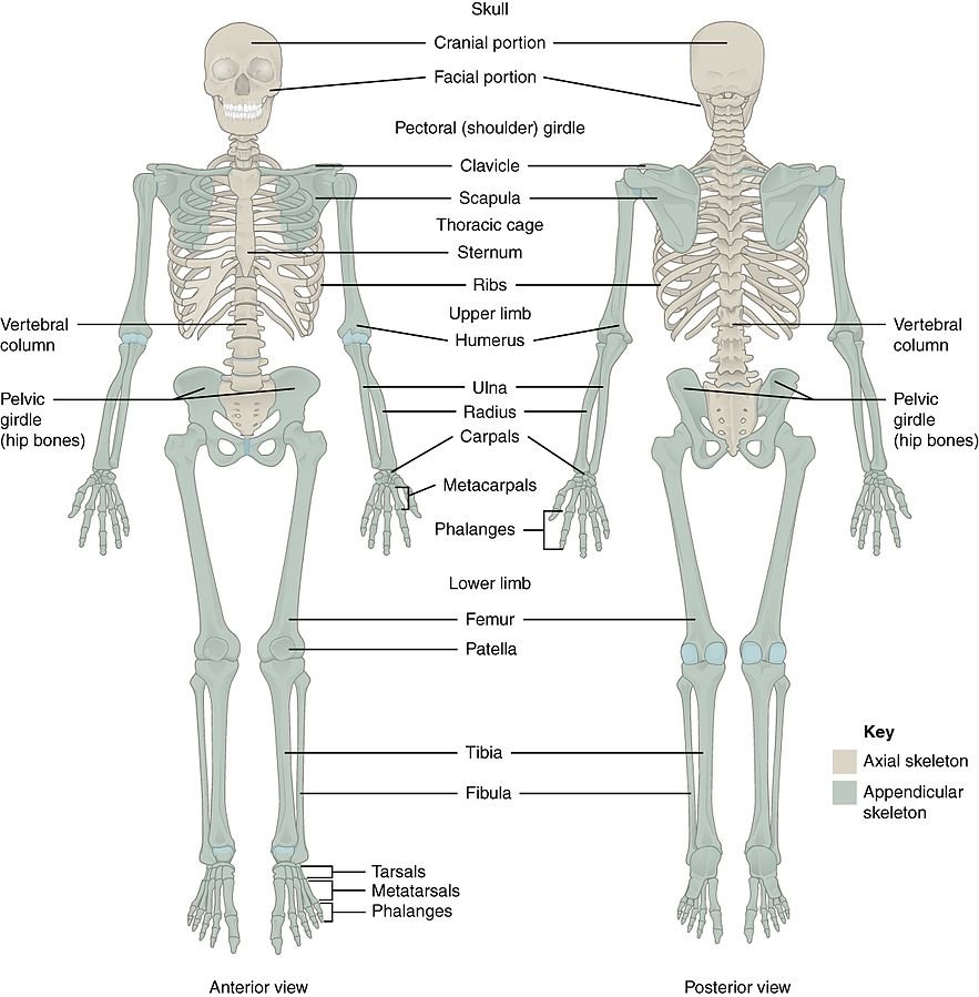
Axial Skeleton
The axial skeleton forms the vertical, central axis of the body and includes all bones of the head, neck, chest, and back. It serves to protect the brain, spinal cord, heart, and lungs. It also serves as the attachment site for muscles that move the head, neck, and back, and for muscles that act across the shoulder and hip joints to move their corresponding limbs.
- Skull: The skull is formed by 22 bones. It consists of two sets of bones: eight cranial bones and 14 facial bones. The cranial bones make up the top and back of your skull and support and protect your brain1. The facial bones make up the face of your skull and form an entrance to your body.
- Vertebral Column: The vertebral column consists of 24 bones, each called a vertebra, plus the sacrum and coccyx. It forms the spine and supports the body, allowing us to stand upright.
- Thoracic Cage: The thoracic cage includes 12 pairs of ribs, and the sternum. It forms the rib cage, which protects the heart and lungs.
- Hyoid Bone and Bones of the Inner Ear: The hyoid bone is located in the throat and does not connect with any other bone, serving as a movable base for the tongue. Inside the temporal bone of the skull are the three smallest bones of the body: the malleus, incus, and stapes, which are located in the middle ear and are responsible for transmitting sound vibrations.
- Number of Bones: The axial skeleton is made up of 80 bones.
Appendicular Skeleton
The appendicular skeleton is one of the two major groups of bones in the human skeleton. It consists of the bones of the limbs (or appendages), and the bones that attach the limbs to the rest of the body. It includes a total of 126 bones, including those in the arms, legs, and shoulder and pelvic girdle bones.
- Shoulder (Pectoral) Girdle: The pectoral or shoulder girdle consists of the scapulae and clavicles. The shoulder girdle connects the bones of the upper limbs to the axial skeleton. These bones also provide attachment for muscles that move the shoulders and upper limbs.
- Bones of the Upper Limbs: The upper limbs include the bones of the arm (humerus), forearm (radius and ulna), wrist, and hand. The only bone of the arm is the humerus, which articulates with the forearm bones–the radius and ulna–at the elbow joint. The wrist, or carpus, consists of eight carpal bones. The hand includes 8 bones in the wrist, 5 bones that form the palm, and 14 bones that form the fingers and thumb.
- Pelvic Girdle: The pelvic girdle is a ring of bones attached to the vertebral column that connects the bones of the lower limbs to the axial skeleton. The pelvic girdle consists of the right and left hip bones. Each hip bone is a large, flattened, and irregularly shaped fusion of three bones: the ilium, ischium, and pubis.
- Bones of the Lower Limbs: The lower limbs include the bones of the thigh, leg, and foot. The femur is the only bone of the thigh. It articulates with the two bones of the leg–the larger tibia (commonly known as the shin) and smaller fibula.
Types of bone
Long Bones
These are bones that are longer than they are wide. They consist of a long shaft with two bulky ends or extremities. They are mostly compact or dense. Examples include the femur, tibia, and fibula in the legs, and the humerus, radius, and ulna in the arms.
Short Bones
These are approximately equal in length, width, and thickness. They are primarily spongy bone, with a thin outer layer of compact bone. Examples include the carpal bones in the wrist and the tarsal bones in the feet.
Flat Bones
These bones are thin, but are often curved. They have a flat shape, not rounded. Examples include certain skull bones, the ribs, the sternum (breastbone), and the scapula (shoulder blade).
Irregular Bones
These bones do not fit into the other bone categories and have complex shapes. Examples include the vertebrae, sacrum, and certain facial bones.
Sesamoid Bones
These are small, round bones that are embedded in tendons. They are located where a tendon passes over a joint, such as the hand, knee, and foot. The patella (kneecap) is the largest sesamoid bone in the body.
Organisation of skeletal muscle
Each skeletal muscle is an organ that consists of various integrated tissues. These tissues include the skeletal muscle fibers, blood vessels, nerve fibers, and connective tissue.
Connective Tissue Layers
Each skeletal muscle has three layers of connective tissue (called “mysia”) that enclose it and provide structure to the muscle as a whole, and also compartmentalize the muscle fibers within the muscle.
- Epimysium: Each muscle is wrapped in a sheath of dense, irregular connective tissue called the epimysium, which allows a muscle to contract and move powerfully while maintaining its structural integrity. The epimysium also separates muscle from other tissues and organs in the area, allowing the muscle to move independently.
- Perimysium: Inside each skeletal muscle, muscle fibers are organized into individual bundles, each called a fascicle, by a middle layer of connective tissue called the perimysium. This fascicular organization is common in muscles of the limbs; it allows the nervous system to trigger a specific movement of a muscle by activating a subset of muscle fibers within a bundle, or fascicle of the muscle.
- Endomysium: Muscle fibers are covered by the endomysium. The endomysium surrounds the extracellular matrix of the cells and plays a role in transferring force produced by the muscle fibers to the tendons.
Muscle Fibers: Muscle fibers are the individual muscle cells within each fascicle. Each muscle fiber is a single cell that contracts in response to stimulation and then relaxes when the stimulation ends.
Fascicles: Fascicles are bundles of muscle fibers. The arrangement of fascicles determines the range of motion and power of a muscle. Inside each fascicle, each muscle fiber is encased in a thin connective tissue layer of collagen and reticular fibers called the endomysium.
Tendons: In skeletal muscles that work with tendons to pull on bones, the collagen in the three connective tissue layers intertwines with the collagen of a tendon. At the other end of the tendon, it fuses with the periosteum coating the bone. The tension created by contraction of the muscle fibers is then transferred though the connective tissue layers, to the tendon, and then to the periosteum to pull on the bone for movement of the skeleton.
Blood Supply: Every skeletal muscle is also richly supplied by blood vessels for nourishment, oxygen delivery, and waste removal.
Nerve Supply: In addition, every muscle fiber in a skeletal muscle is supplied by the axon branch of a somatic motor neuron, which signals the fiber to contract.
Neuromuscular junction
A neuromuscular junction (or myoneural junction) is a chemical synapse between a motor neuron and a muscle fiber. It allows the motor neuron to transmit a signal to the muscle fiber, causing muscle contraction.
Components
Each muscle fiber is innervated (supplied) and controlled by a motor neuron. This motor neuron, which has its cell body located within the central nervous system, will have axons that enter the muscle and penetrate the perimysium. At this point, each axon of the motor neuron will divide into branches called axon terminals. The synaptic end bulb of the motor neuron comprises the nervous system component of the neuromuscular junction. The muscular component is a region of the muscle fiber referred to as the motor end plate. Between the synaptic end bulbs of the neuron and the cell membrane of the muscle fiber (the sarcolemma) lies a space known as the synaptic cleft, which is the final component of the neuromuscular junction.
Synaptic Transmission
Synaptic transmission at the neuromuscular junction begins when an action potential reaches the presynaptic terminal of a motor neuron, which activates voltage-gated calcium channels to allow calcium ions to enter the neuron. Calcium ions bind to sensor proteins (synaptotagmins) on synaptic vesicles, triggering vesicle fusion with the cell membrane and subsequent neurotransmitter release from the motor neuron into the synaptic cleft. In vertebrates, motor neurons release acetylcholine (ACh), a small molecule neurotransmitter, which diffuses across the synaptic cleft and binds to nicotinic acetylcholine receptors (nAChRs) on the cell membrane of the muscle fiber, also known as the sarcolemma1. nAChRs are ionotropic receptors, meaning they serve as ligand-gated ion channels. The binding of ACh to the receptor can depolarize the muscle fiber, causing a cascade that eventually results in muscle contraction.
Physiology of muscle contraction
Neuromuscular Junction
The process of muscle contraction begins at the site where a motor neuron’s terminal meets the muscle fiber—called the neuromuscular junction (NMJ). Every skeletal muscle fiber in every skeletal muscle is innervated by a motor neuron at a NMJ. Excitation signals from the motor neuron are the only way to functionally activate skeletal muscle fibers to contract.
Excitation-Contraction Coupling
All living cells have membrane potentials, or electrical gradients across their membranes based on the distribution of positively and negatively charged ions. Neurons and muscle cells can use their membrane potentials to generate and conduct electrical signals by controlling the movement of charged ions across their membranes to create electrical currents. This movement is controlled by selective opening and closing of specialized proteins in the membrane called ion channels. Although the currents generated by ions moving through these channel proteins are very small, they form the basis of both neural signaling and muscle contraction.
Action Potentials
Both neurons and skeletal muscle cells are electrically excitable, meaning that they are able to generate action potentials. An action potential is a special type of electrical signal that can travel along a cell membrane as a wave. This allows a signal to be transmitted quickly over long distances.
Muscle Contraction
In skeletal muscle, cross-bridge formation and contraction requires the presence of calcium (Ca ++) inside the muscle cell. Excitation signaling of action potentials from the motor neuron are coupled with calcium release. Thus, the excitation-contraction coupling process begins with signaling from the nervous system at the neuromuscular junction and ends with calcium release for muscle contraction.
Cross-Bridge Cycling
The whole process is called the mechanism of muscle contraction and it can be summarized in three steps:
- A message travels from the nervous system to the muscular system, triggering chemical reactions.
- The chemical reactions lead to the muscle fibers reorganizing themselves in a way that shortens the muscle–that’s the contraction.
- When the nervous system signal is no longer present, the chemical process reverses, and the muscle fibers rearrange again and the muscle relaxes.
List axial skeletal system bones
- Skull: 22 bones
- Cranial Bones: 8 (Frontal Bone, 2 Parietal Bones, 2 Temporal Bones, Occipital Bone, Ethmoid Bone, Sphenoid Bone)
- Facial Bones: 14 (2 Maxillae, 2 Lacrimal Bones, 2 Zygomatic Bones, 2 Palatine Bones, 2 Nasal Bones, 2 Inferior Nasal Conchae, Vomer, Mandible)
- Ossicles of Middle Ear: 6 bones (3 in each ear: Malleus, Incus, Stapes)
- Hyoid Bone: 1 bone
- Vertebral Column: 26 bones
- Cervical Vertebrae: 7
- Thoracic Vertebrae: 12
- Lumbar Vertebrae: 5
- Sacrum: 1 (fused from 5 sacral vertebrae)
- Coccyx: 1 (fused from 4 coccygeal vertebrae)
- Thoracic Cage: 25 bones
- Ribs: 24 (12 pairs)
- Sternum: 1
So, the axial skeleton is made up of 80 bones.
List appendicular skeletal system bones
- Shoulder (Pectoral) Girdle: 4 bones (2 Clavicles, 2 Scapulae)
- Upper Limbs: 60 bones
- Arms: 2 Humeri
- Forearms: 2 Radii, 2 Ulnae
- Hands: 54 (16 Carpals, 10 Metacarpals, 28 Phalanges)
- Pelvic Girdle: 2 bones (2 Hip Bones)
- Lower Limbs: 60 bones
- Thighs: 2 Femurs
- Legs: 2 Tibiae, 2 Fibulae
- Feet: 56 (14 Tarsals, 10 Metatarsals, 28 Phalanges, 4 Sesamoid)
The appendicular skeletal system consists of a total of 126 bones.
Conclusion
The skeletal system, comprising the axial and appendicular skeletons, is a marvel of biological engineering. With a total of 206 bones in an adult human body, it provides not only the structural framework that supports and gives shape to the body, but also plays crucial roles in protection, movement, and blood cell production.
The axial skeleton, consisting of 80 bones, forms the central core of the body, providing vital protection for our brain, spinal cord, heart, and lungs. The appendicular skeleton, on the other hand, consists of 126 bones and includes the bones of our limbs and the girdles that attach these limbs to the axial skeleton.
Each bone, whether it’s the tiny stapes in our ear or the large femur in our thigh, has a specific structure and function, contributing to our body’s overall health and movement. Understanding the intricacies of the skeletal system is not only fascinating but also crucial for fields ranging from medicine to anthropology.
For more regular updates you can visit our social media accounts,
Instagram: Follow us
Facebook: Follow us
WhatsApp: Join us
Telegram: Join us
2 thoughts on “Skeletal System”