Special senses- Eye
Special senses-eye, a marvel of biological engineering, is our window to the world. It is an intricate organ that captures light, processes it into images, and sends these signals to the brain, enabling us to perceive our surroundings. The eye’s complexity is matched only by its importance, as vision is often considered our most valued sense. This article delves into the fascinating world of the human eye, exploring its anatomy, common diseases, and tips for maintaining optimal eye health.
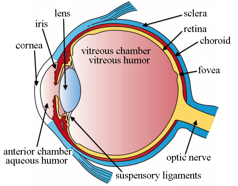
Structure of eye
Orbit
- Structure: The orbit is a paired, conical or four-sided pyramidal cavity within the skull, each comprising seven bones. These bones range from the paper-thin ethmoid and lacrimal plate medially to the buttress thick zygoma laterally.
- Function: The primary function of the orbit is to protect and accommodate the globe (eye) in order to maximize its function. It provides mechanical protection for the eye and soft tissue structures related to it.
- Composition: The orbit is made up of several bones. The medial wall comprises the ethmoid bone in the center, the lacrimal bone and maxilla – or maxillary bone anteriorly, and the lesser wing of the sphenoid bone posteriorly. The superior wall – or roof of the orbit – is mainly formed by the orbital part of the frontal bone anteriorly, and a small posterior part by the lesser wing of the sphenoid bone. The lateral wall is made up of the zygomatic bone anteriorly and the greater wing of the sphenoid bone posteriorly. Finally, the inferior wall – or floor of the orbit – is formed by the maxillary bone medially, the zygomatic bone laterally, and a tiny part by the palatine bone posteriorly.
- Protection: The orbit also protects the muscles, vessels, and nerves of the eyes. Between each eye and the orbit protecting it, there’s a soft cushion of fat to prevent any friction or damage to the eyes.
- Foramina and Fissures: The bones that make up the orbit contain several foramina and fissures through which important neurovascular structures (such as the optic nerve (CN II)) pass through on their way from the brain to the eye and face and vice versa.
Sclera
- Structure and Location: The sclera is the white part of your eye. It’s a protective covering that wraps over most of your eyeball. It extends from the cornea at the front of your eye to the optic nerve in the back.
- Composition: The sclera is made of tough collagen fibers. Collagen is a protein that’s the main ingredient in your body’s skin, muscles, bones and connective tissues. The collagen fibers in your sclera crisscross each other in a random pattern that makes the sclera strong and flexible.
- Function: The sclera is the supporting wall for your eyeball. It maintains your eye’s shape and protects it from injuries. Muscles attached to the sclera help you move your eyeball.
- Protection: The sclera acts as a protective layer for the delicate internal structures of the eye, including the retina, choroid, and optic nerve1. It helps shield the eye from external physical injuries and provides a barrier against dust, debris, and other environmental elements.
- Maintenance of Intraocular Pressure: The sclera, along with the cornea (the transparent front part of the eye), helps maintain intraocular pressure. This pressure is essential for the proper functioning of the eye and is necessary for maintaining the shape of the eyeball.
- Attachment for Eye Muscles: Six muscles control the movement of each eye. The extrinsic eye muscles responsible for eye movement attach to the sclera. These muscles allow the eyes to move in different directions, facilitating activities such as tracking moving objects and maintaining proper alignment of the eyes.
Conjunctiva
- Structure and Location: The conjunctiva is a thin, clear membrane that covers the white part of the eye (sclera) and lines the inside of the eyelids. It is continuous with the skin of the eyelids and with the lacrimal canaliculi and lacrimal sac.
- Composition: The conjunctiva is made up of cells and tissues that have specific jobs. For example, goblet cells secrete mucus and a layer of cells called the stratified squamous epithelium provides structural stability to the eye. The conjunctiva is made of three parts: the palpebra conjunctiva (the lining of your eyelids), the bulbar conjunctiva (the covering over your eyeball that protects the white of your eye), and the conjunctiva fornix (a piece that joins the palpebra and bulbar conjunctiva together where they meet).
- Function: The main function of the conjunctiva is to keep the eye moist. It provides lubrication to the eyes by making mucus and tears. These fluids form a covering (tear film) that has three layers: innermost mucus layer, the middle watery layer, and the outer oily layer. The tear film has a few important jobs: barrier protection, lubrication, visual acuity, and eye health.
- Protection: The conjunctiva acts like a combination of a raincoat and plastic wrap. It blocks irritants (like allergens) from getting into your eye and keeps moisture and lubrication sealed in. You might develop dry eyes or an eye infection if something’s wrong with your conjunctiva.
Tear Film
Structure: The tear film is a thin fluid layer that covers the ocular surface. It is traditionally described as being divided into three layers:
- Oily Layer: This is the outermost layer of the tear film. It is produced by the meibomian glands and helps prevent the tears from drying up too quickly. It also makes the surface of the eyes smooth.
- Watery Layer: This is the middle layer of the tear film, which is produced by the lacrimal glands. It keeps the eyes wet and nourishes the eye tissue.
- Mucin Layer: This is the innermost layer of the tear film, which is produced by the goblet cells of the conjunctiva. It helps the tear film stick to the surface of the eyes.
Function: The tear film serves several important functions:
- Lubrication: It provides ocular surface comfort through continuous lubrication.
- Protection: The tear film serves to protect against irritants, allergens, environmental extremes of dryness and temperature, potential pathogens, and pollutants.
- Nourishment: The tear film supplies glucose, electrolytes, and growth factors to the cornea, which is an avascular structure.
- Vision: The tear film provides a smooth and very powerful refracting surface for clear vision.
- Production and Regulation: Tears are continually replenished from the inferior tear meniscus by blinking. This counters the forces of gravity and evaporation on the volume of the precorneal tear film and protects corneal and conjunctival epithelial cells from the shear forces exerted by the eyelids during blinking.
Cornea
- Location and Structure: The cornea is the clear, dome-shaped front part of the eye. It covers the pupil (the opening at the center of the eye), iris (the colored part of the eye), and anterior chamber (the fluid-filled inside of the eye). The cornea forms the anterior transparent layer of the outer coat of the eye.
- Composition: The cornea is composed of proteins and cells. It does not contain blood vessels, unlike most tissues in the human body. It consists of five layers: the corneal epithelium, anterior limiting lamina (Bowman’s layer), substantia propria (stroma), posterior limiting lamina (Descemet’s membrane), and endothelium.
- Function: The cornea’s main function is to refract, or bend, light. It acts as the eye’s outermost lens and controls and focuses the entry of light into the eye. The cornea contributes between 65-75 percent of the eye’s total focusing power. When light strikes the cornea, it bends–or refracts–the incoming light onto the lens.
- Protection: The cornea contributes to the protection of the eye by creating a structural barrier that protects the eye from bacteria and subsequent infections.
- Nutrient Supply: Since there are no nutrient-supplying blood vessels in the cornea, tears and the aqueous humor (a watery fluid) in the anterior chamber provide the cornea with nutrients.
Anterior Chamber
- Structure and Location: The anterior chamber is one of three fluid-filled spaces in the eye. It is located between the iris (the colored part of the eye) and the cornea’s innermost surface, the endothelium.
- Composition: The anterior chamber is filled with a fluid called the aqueous humor. This clear, watery fluid nourishes the internal structures of the eye.
- Function: The anterior chamber allows the aqueous humor to flow to the eye to carry out vital functions. The fluid flows from the posterior chamber, through the pupil, and into the anterior chamber. From there, it drains out of the eye through structures in the angle of the anterior chamber
Iris
- Structure and Location: The iris is the colored part of the eye. It is part of the vascular layer of the eye, which also includes the choroid and the ciliary body. The central opening of the iris is the pupil.
- Composition: The iris is a flat and ring-shaped membrane behind the cornea of the eye. Within the iris, there are smooth muscle fibers that control the size of the pupil. These muscle fibers form the sphincter pupillae muscle, which constricts the pupillary opening, and the dilator pupillae muscle, which dilates the pupil.
- Function: The iris controls the size of the pupils and thus regulates the amount of light entering the eye. It is the structure that provides an individual with eye color.
Pupil
- Structure and Location: The pupil is the small black circle located in the center of the eyeball. Surrounding the pupil is the colored part of the eye, the iris.
- Function: The pupil’s function is to allow light to pass through and enter the eye. This light then interacts with the cells of the retina, working as part of the visual pathway to provide the ability of sight. The size of the pupil is controlled by muscles within the iris — one muscle constricts the pupil opening (makes it smaller), and another iris muscle dilates the pupil (makes it larger).
- Size: The size of the pupil varies from person to person. Some people have large pupils, and some people have small pupils. Also, pupil size changes with age — children and young adults tend to have large pupils, and seniors usually have small pupils4. Generally, normal pupil size in adults ranges from 2 to 4 millimeters (mm) in diameter in bright light to 4 to 8 mm in the dark.
Lens
- Structure and Location: The lens is an ellipsoid structure located in the eyeball. It lies posterior to the iris and anterior to the vitreous body. Moreover, the lens is encircled by the ciliary processes, and is attached to them by the zonular fibres.
- Composition: The lens is comprised of three main parts: capsule, epithelium, and fibers. Although it has many layers, the lens is transparent. The crystalline lens is a clear, biconvex layer of the eye that is made up mostly of proteins. As much as 60% of the lens mass is made up of proteins—a concentration higher than almost any other tissue in the body.
- Function: The main function of the lens is to transmit and focus the light onto the retina in order to create clear images of observed objects at various distances. The lens is also the main structure of the accommodation reflex. This reflex is activated when the eye focuses on closer objects. The activation of the reflex pathway leads to the increase of the lens curvature which increases the refraction power of the eye (i.e. eye’s focus).
- Protection and Nourishment: The lens absorbs ultraviolet (UV) light of less than 350 nm wavelength, thus preventing damaging UV radiation from reaching the retina. The lens has no direct blood or nerve connections. Instead, it relies on the aqueous humor—the clear fluid between the lens and the cornea—to provide it with energy and carry away waste products.
Vitreous Cavity
- Structure and Location: The vitreous cavity is the largest of the three fluid-filled spaces in the eye. It is located behind the lens and in front of the optic nerve. This chamber is filled with a thick, clear gel-like substance called the vitreous humor (also known as vitreous body).
- Composition: The vitreous humor is composed of mostly water, along with a small percentage of collagen, glycosaminoglycans (sugars), electrolytes (salts), and proteins. The vitreous humor is transparent, and functions in transporting nutrients to the lens, ciliary body and retina.
- Function: The vitreous humor’s main role is to maintain the round shape of the eye. The size and shape of the vitreous humor also ensures that it remains attached to the retina, which is the layer at the back of the eye that is sensitive to light. The vitreous humor is also a part of the eye that can help with vision clarity. Because the vitreous humor is a clear substance, light is able to pass through and reach the retina. Near the center of the retina is the macula, a pigmented region responsible for high-resolution color vision.
- Protection: The vitreous humor can also be helpful in absorbing any unexpected disturbances to the eye, such as a thump to the side of head. Absorbing the shock associated with the thump or similar disturbance can help prevent eye damage.
- Changes Over Time: With the normal process of aging, the vitreous humor may begin to shrink due to a decrease in viscosity or thickness. This process is called vitreous degeneration. As the fluid changes from a thick gel-like substance to a thinner liquid consistency, the vitreous humor separates from the retina. This can lead to vitreous floaters, or small disruptions in the visual field such as spots, web-like lines, or rings.
Retina
- Structure and Location: The retina is the innermost layer of the eye. It starts at the posterior surface of the eyeball and terminates anteriorly at the ora serrata. It is found between the choroid and vitreous body.
- Composition: The retina is composed of epithelial, glial, and neural cells that are organized into 10 distinctive layers. Out of these, the first 9 layers belong to the inner neurosensory retina, one of which are the photoreceptors that are sensitive to light. The 10th layer constitutes the outer retinal pigmented epithelium (RPE), which serves to absorb light that passes through the retina and prevent it from reflecting back to the neurosensory layer.
- Function: The function of the retina is to convert visual stimuli from the outside environment into neural impulses that are transmitted to the cerebral cortex via the optic nerve for interpretation and analysis1. The retina senses light and generates electrical impulses so the brain can create an image.
- Photoreceptors: The retina contains millions of light-sensitive cells called photoreceptors. These specialized cells are responsible for turning light into signals that are sent to the brain. There are two main types of photoreceptors: Rod cells, which are extremely sensitive to light and help you see in dim light conditions, and Cone cells, which provide color vision and help you see fine details.
- Macula and Fovea: The macula is the retina’s center, where cone cells are concentrated. This part of the eye helps you see details in the center of your visual field. The fovea is the very center of the macula, where vision is the sharpest.
- Peripheral Retina: This is the area of the retina outside the macula, responsible for peripheral vision and night vision. Rod cells are more abundant in the peripheral retina.
Functions of eye
- Light Reception: The primary function of the eye is to receive light. The cornea and lens work together to focus incoming light onto the retina.
- Image Formation: The cornea, a dome-shaped structure, bends the light to help the eye focus. The iris controls the amount of light that enters the eye through the pupil.
- Adjustment to Light Intensity: The iris, the colored part of the eye, adjusts the size of the pupil to control the amount of light entering the eye. In bright conditions, the iris constricts the pupil to let in less light, and in dark conditions, it dilates the pupil to let in more light.
- Focusing: The lens, located behind the iris and pupil, changes its shape to focus light onto the retina. It becomes thicker to focus on nearby objects and thinner to focus on distant objects.
- Conversion to Electrical Signals: The retina contains photoreceptor cells (rods and cones) that convert light into electrical signals.
- Color and Light Perception: The cones in the retina help perceive color and detail, while the rods help with low-light and peripheral vision.
- Signal Transmission: The optic nerve carries the electrical signals from the retina to the brain, where they are processed into the images we see.
- Protection: The eyelids, eyelashes, and tear film work together to protect the eye from dust, foreign particles, and harsh environmental conditions.
- Lubrication: The tear film also keeps the eye moist and lubricated, which is essential for clear vision and comfort.
- Movement: The extraocular muscles allow the eye to move in various directions, enabling us to look around without moving our heads.
Disorders of eye
- Macular Degeneration: This is primarily an age-related retinal condition. There are two types of macular degeneration — wet and dry. You may lose central vision, but you aren’t likely to lose all of your vision. There are treatments, but there isn’t a cure.
- Cataract: A cataract is a clouding of the lens of the eye, which is typically clear. For people who have cataracts, seeing through cloudy lenses is like looking through a frosty or fogged-up window. Clouded vision caused by cataracts can make it more difficult to read, drive a car at night or see the expression on a friend’s face.
- Glaucoma: Glaucoma is an eye disease that damages your eye’s optic nerve. It usually happens when fluid builds up in the front part of your eye. That extra fluid increases the pressure in your eye, damaging the optic nerve.
- Retinal Detachment: Retinal detachment is a disorder of the eye in which the retina peels away from its underlying layer of support tissue. Initial detachment may be localized, but without rapid treatment the entire retina may detach, leading to vision loss and blindness.
- Amblyopia (Lazy Eye): Amblyopia is a condition in which one eye cannot focus as clearly as the other, which is characterized by decreased vision.
- Strabismus (Crossed Eyes): Strabismus is misalignment of the eyes, causing one eye to deviate inward (esotropia) toward the nose, or outward (exotropia), while the other eye remains focused.
- Conjunctivitis (Pink Eye): Conjunctivitis is an inflammation of the transparent covering of the eye because of bacterial or viral infection or allergic reaction. The eye appears swollen, and red with itching sensation.
- Presbyopia: Presbyopia is the loss of near vision that happens with aging due to hardening of the lens. Symptoms include blurry close-up vision, headaches and eye strain.
- Diabetes-related Retinopathy: In diabetes, elevated blood sugar levels damage the blood vessels of the retina. These damaged blood vessels leak fluid, bleed, and do not provide adequate oxygen to the retina, leading to retinal ischemia.
Eye Care Tips
Maintaining eye health is crucial for preserving our vision. Here are some tips for keeping your eyes in top shape:
- Eat Well: A balanced diet rich in fruits, vegetables, and omega-3 fatty acids can help protect your sight.
- Quit Smoking: Smoking increases the risk of cataracts, optic nerve damage, and macular degeneration.
- Wear Sunglasses: Protect your eyes from the sun’s ultraviolet rays to reduce the risk of cataracts and macular degeneration.
- Use Safety Eyewear: Protect your eyes from injury at home or work by using safety glasses or goggles.
- Take Breaks: Rest your eyes regularly, especially when reading, working at a computer, or driving long distances.
- Regular Check-ups: Regular comprehensive dilated eye exams can help detect eye diseases ear.
Conclusion
In conclusion, the human eye is a marvel of biological engineering, a complex organ that plays a crucial role in our perception of the world. From the protective sclera and conjunctiva to the light-sensitive retina, each part of the eye performs a unique function that contributes to our vision. The cornea, iris, and lens work together to focus light onto the retina, which then sends signals to the brain to be interpreted as images. The vitreous cavity and anterior chamber maintain the shape of the eye and nourish its internal structures. The tear film keeps the eye moist and lubricated, ensuring comfort and clear vision. However, the eye is susceptible to various diseases and conditions, emphasizing the importance of regular check-ups and proper eye care. As we continue to unravel the mysteries of this remarkable organ, we gain a deeper appreciation for the gift of sight and the intricate mechanisms that make it possible.
For more regular updates you can visit our social media accounts,
Instagram: Follow us
Facebook: Follow us
WhatsApp: Join us
Telegram: Join us
2 thoughts on “Special Senses- Eye”